TUBA4A Primary Antibody

Item Information
Catalog #
Size
Price
Description

Microtubules of the eukaryotic cytoskeleton perform essential and diverse functions and are composed of a heterodimer of alpha and beta tubulin. The genes encoding these microtubule constituents are part of the tubulin superfamily, which is composed of six distinct families. Genes from the alpha, beta and gamma tubulin families are found in all eukaryotes. The alpha and beta tubulins represent the major components of microtubules, while gamma tubulin plays a critical role in the nucleation of microtubule assembly. There are multiple alpha and beta tubulin genes and they are highly conserved among and between species. This gene encodes an alpha tubulin that is a highly conserved homolog of a rat testis-specific alpha tubulin. Alternatively spliced transcript variants encoding different isoforms have been found for this gene. [provided by RefSeq, Jun 2013]
Product Overview
Entrez GenelD
7277
Aliases
ALS22; TUBA1; H2-ALPHA
Clone#
7B11E6
Host / Isotype
Mouse / Mouse IgG1
Immunogen
Purified recombinant fragment of human TUBA4A (AA: (299-447)) expressed in E. Coli.
Formulation
Purified antibody in PBS with 0.05% sodium azide
Storage
4°C; -20°C for long term storage
Product Applications
WB (Western Blot)
1/500 - 1/2000
IHC_P(Immunohistochemistry)
1/200-1/1000
FCM (Flow Cytometry)
1/200-1/400
ELISA
1/10000
References
1,J Neurol Neurosurg Psychiatry. 2018 Dec;89(12):1350-1352.2,Neurodegener Dis. 2017;17(4-5):171-180.
Product Image
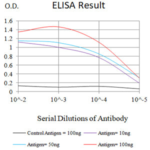
ELISA
Figure 1: Black line: Control Antigen (100 ng);Purple line: Antigen (10ng); Blue line: Antigen (50 ng); Red line: Antigen (100 ng)
WESTERN BLOT
Figure 2: Western blot analysis using TUBA4A mAb against human TUBA4A (AA: (299-447)) recombinant protein. (Expected MW is 36.9 kDa)
WESTERN BLOT
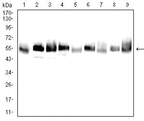
Figure 3: Western blot analysis using TUBA4A mouse mAb against A431 (1), Hela (2), HepG2 (3), Jurkat (4), Cos7 (5),C6 (6), NIH3T3 (7), HEK293 (8),and HEK293-6e (9) cell lysate.
FLOW CYTOMETRY
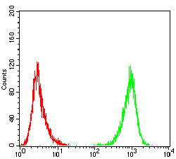
Figure 4: Flow cytometric analysis of A431 cells using TUBA4A mouse mAb (green) and negative control (red).
FLOW CYTOMETRY
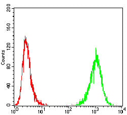
Figure 5: Flow cytometric analysis of Hepg2 cells using TUBA4A mouse mAb (green) and negative control (red).
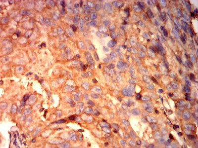
IMMUNOHISTOCHEMISTRY
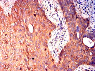
Figure 6: Immunohistochemical analysis of paraffin-embedded cervical cancer tissues using TUBA4A mouse mAb with DAB staining.
IMMUNOHISTOCHEMISTRY
Figure 7: Immunohistochemical analysis of paraffin-embedded ovarian cancer tissues using TUBA4A mouse mAb with DAB staining.
For Research Use Only. Not for use in diagnostic procedures.