SMC1 Primary Antibody

Item Information
Catalog #
Size
Price
Description

Proper cohesion of sister chromatids is a prerequisite for the correct segregation of chromosomes during cell division. The cohesin multiprotein complex is required for sister chromatid cohesion. This complex is composed partly of two structural maintenance of chromosomes (SMC) proteins, SMC3 and either SMC1L2 or the protein encoded by this gene. Most of the cohesin complexes dissociate from the chromosomes before mitosis, although those complexes at the kinetochore remain. Therefore, the encoded protein is thought to be an important part of functional kinetochores. In addition, this protein interacts with BRCA1 and is phosphorylated by ATM, indicating a potential role for this protein in DNA repair. This gene, which belongs to the SMC gene family, is located in an area of the X-chromosome that escapes X inactivation.

Product Overview
Entrez GenelD
8243
Aliases
SMC1; SMCB; CDLS2; SB1.8; SMC1L1; DXS423E; KIAA0178; MGC138332; SMC1alpha; DKFZp686L19178; SMC1A
Clone#
5B6
Host / Isotype
Mouse / IgG1
Species Reactivity
Human
Immunogen
Purified recombinant fragment of human SMC1 expressed in E. Coli.
Formulation
Purified antibody in PBS with 0.05% sodium azide
Storage
4°C; -20°C for long term storage
Product Applications
WB (Western Blot)
1/500 - 1/2000
IHC_P(Immunohistochemistry)
1/200 - 1/1000
ICC (Immunocytochemistry)
1/200 - 1/1000
FCM (Flow Cytometry)
1/200 - 1/400
ELISA
1/10000
References
1. Cell Cycle. 2006 Nov 1;5(21):2537-42.
2. FEBS Lett. 2007 Jun 26;581(16):3005-12.
Product Image
Western Blot
Figure 1: Western blot analysis using SMC1 mouse mAb against K562 (1), Jurkat (2) and A549 (3) cell lysate.
Immunohistochemical analysis
Figure 2: Immunohistochemical analysis of paraffin-embedded human colon using SMC1 mouse mAb with DAB staining.
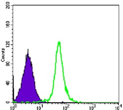
Flow cytometric
Figure 3: Flow cytometric analysis of Hela cells using SMC1 mouse mAb (green) and negative control (purple).
Immunofluorescence analysis
Figure 4: Immunofluorescence analysis of NIH/3T3 cells using SMC1 mouse mAb (green). Red: Actin filaments have been labeled with Alexa Fluor-555 phalloidin.
For Research Use Only. Not for use in diagnostic procedures.