SMAD2 Primary Antibody

Item Information
Catalog #
Size
Price
Description

The protein encoded by this gene belongs to the SMAD, a family of proteins similar to the gene products of the Drosophila gene 'mothers against decapentaplegic' (Mad) and the C. elegans gene Sma. SMAD proteins are signal transducers and transcriptional modulators that mediate multiple signaling pathways. This protein mediates the signal of the transforming growth factor (TGF)-beta, and thus regulates multiple cellular processes, such as cell proliferation, apoptosis, and differentiation. This protein is recruited to the TGF-beta receptors through its interaction with the SMAD anchor for receptor activation (SARA) protein. In response to TGF-beta signal, this protein is phosphorylated by the TGF-beta receptors. The phosphorylation induces the dissociation of this protein with SARA and the association with the family member SMAD4. The association with SMAD4 is important for the translocation of this protein into the nucleus, where it binds to target promoters and forms a transcription repressor complex with other cofactors. This protein can also be phosphorylated by activin type 1 receptor kinase, and mediates the signal from the activin. Alternatively spliced transcript variants encoding the same protein have been observed.

Product Overview
Entrez GenelD
4087
Aliases
JV18; MADH2; MADR2; JV18-1; hMAD-2; hSMAD2; MGC22139; MGC34440
Clone#
5G7
Host / Isotype
Mouse / IgG1
Species Reactivity
Human
Immunogen
Purified recombinant fragment of human SMAD2 expressed in E. Coli.
Formulation
Ascitic fluid containing 0.03% sodium azide.
Storage
4°C; -20°C for long term storage
Product Applications
WB (Western Blot)
1/500 - 1/2000
IHC_P(Immunohistochemistry)
1/200 - 1/1000
ICC (Immunocytochemistry)
1/200 - 1/1000
FCM (Flow Cytometry)
1/200 - 1/400
ELISA
1/10000
References
1. J Biol Chem. 2009 Dec 4;284(49):34145-56.
2. Cloning Stem Cells. 2009 Sep;11(3):427-35.
Product Image
Western Blot
Figure 1: Western blot analysis using SMAD2 mAb against human SMAD2 (AA: 20-254) recombinant protein. (Expected MW is 52.2 kDa)
Western Blot
Figure 2: Western blot analysis using SMAD2 mAb against HEK293 (1) and SMAD2(AA: 20-254)-hIgGFc transfected HEK293 (2) cell lysate.
Immunohistochemical analysis
Figure 3: Immunohistochemical analysis of paraffin-embedded human liver cancer tissues using SMAD2 mouse mAb with DAB staining.
Immunohistochemical analysis
Figure 4: Immunohistochemical analysis of paraffin-embedded human cerebellum tissues using SMAD2 mouse mAb with DAB staining.
Immunofluorescence analysis
Figure 5: Immunofluorescence analysis of U251 cells using SMAD2 mouse mAb (green). Blue: DRAQ5 fluorescent DNA dye. Red: Actin filaments have been labeled with Alexa Fluor-555 phalloidin.
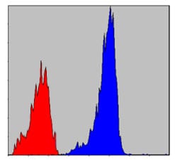
Flow cytometric
Figure 6: Flow cytometric analysis of NIH/3T3 cells using SMAD2 mouse mAb (blue) and negative control (red).
Elisa
Red: Control Antigen (100ng); Purple: Antigen (10ng); Green: Antigen (50ng); Blue: Antigen (100ng);
For Research Use Only. Not for use in diagnostic procedures.