S100A4
Item Information
Catalog #
Size
Price
Description
The protein encoded by this gene is a member of the S100 family of proteins containing 2 EF-hand calcium-binding motifs. S100 proteins are localized in the cytoplasm and/or nucleus of a wide range of cells, and involved in the regulation of a number of cellular processes such as cell cycle progression and differentiation. S100 genes include at least 13 members which are located as a cluster on chromosome 1q21. This protein may function in motility, invasion, and tubulin polymerization. Chromosomal rearrangements and altered expression of this gene have been implicated in tumor metastasis. Multiple alternatively spliced variants, encoding the same protein, have been identified.
Product Overview
Entrez GenelD
6275
Aliases
42A; 18A2; CAPL; FSP1; MTS1; P9KA; PEL98
Clone#
3A1B2
Host / Isotype
Mouse / Mouse IgG1
Species Reactivity
Human, Mouse, Rat
Immunogen
Purified recombinant fragment of human S100A4 (AA: 2-101) expressed in E. Coli.
Formulation
Purified antibody in PBS with 0.05% sodium azide
Storage
4℃; -20℃ for long term storage
Product Applications
WB (Western Blot)
1/500 - 1/2000
IHC_P(Immunohistochemistry)
1/200 - 1/1000
FCM (Flow Cytometry)
1/200 - 1/400
ELISA
1/10000
References
1.Int J Mol Sci. 2021 Apr 29;22(9):4720.
2.Invest Ophthalmol Vis Sci. 2020 Sep 1;61(11):19.
2.Invest Ophthalmol Vis Sci. 2020 Sep 1;61(11):19.
Product Image
Elisa

Figure 1:Black line: Control Antigen (100 ng);Purple line: Antigen (10ng); Blue line: Antigen (50 ng); Red line:Antigen (100 ng)
Western Blot

Figure 2:Western blot analysis using S100A4 mAb against human S100A4 (AA: 2-101) recombinant protein. (Expected MW is 37.5 kDa)
Western Blot

Figure 3:Western blot analysis using S100A4 mAb against HEK293-6e (1) and S100A4 (AA: 2-101)-hIgGFc transfected HEK293-6e (2) cell lysate.
Immunofluorescence analysis

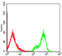
Figure 4:Flow cytometric analysis of Jurkat cells using S100A4 mouse mAb (green) and negative control (red).
Immunohistochemical analysis

Figure 5:Immunohistochemical analysis of paraffin-embedded breast cancer tissues using S100A4 mouse mAb with DAB staining.
Immunohistochemical analysis

Figure 6:Immunohistochemical analysis of paraffin-embedded rectum cancer tissues using S100A4 mouse mAb with DAB staining.
Immunohistochemical analysis

Figure 7:Immunohistochemical analysis of paraffin-embedded mouse spleen tissues using S100A4 mouse mAb with DAB staining.
Immunohistochemical analysis

Figure 8:Immunohistochemical analysis of paraffin-embedded Rat spleen tissues using S100A4 mouse mAb with DAB staining.
Immunohistochemical analysis

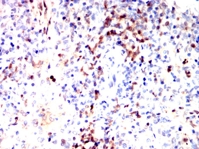
Figure 9:Immunohistochemical analysis of paraffin-embedded Rabbit spleen tissues using S100A4 mouse mAb with DAB staining.
Immunohistochemical analysis

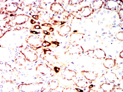
Figure 10:Immunohistochemical analysis of paraffin-embedded Rabbit kidney tissues using S100A4 mouse mAb with DAB staining.
For Research Use Only. Not for use in diagnostic procedures.

