RFA2

Item Information
Catalog #
Size
Price
Description

This gene encodes a subunit of the heterotrimeric Replication Protein A (RPA) complex, which binds to single-stranded DNA (ssDNA), forming a nucleoprotein complex that plays an important role in DNA metabolism, being involved in DNA replication, repair, recombination, telomere maintenance, and co-ordinating the cellular response to DNA damage through activation of the ataxia telangiectasia and Rad3-related protein (ATR) kinase. The RPA complex protects single-stranded DNA from nucleases, prevents formation of secondary structures that would interfere with repair, and co-ordinates the recruitment and departure of different genome maintenance factors. The heterotrimeric complex has two different modes of ssDNA binding, a low-affinity and high-affinity mode, determined by which oligonucleotide/oligosaccharide-binding (OB) domains of the complex are utilized, and differing in the length of DNA bound. This subunit contains a single OB domain that participates in high-affinity DNA binding and also contains a winged helix domain at its carboxy terminus, which interacts with many genome maintenance protein. Post-translational modifications of the RPA complex also plays a role in co-ordinating different damage response pathways.

Product Overview
Entrez GenelD
6118
Aliases
RP-A p34, RPA2, REPA2, RPA32, RPA34
Clone#
2C5B6
Host / Isotype
Mouse / Mouse IgG1,k
Species Reactivity
Human
Immunogen
Purified recombinant fragment of human RFA2 expressed in E. Coli.
Formulation
Purified antibody in PBS with 0.05% sodium azide
Storage
4℃; -20℃ for long term storage
Product Applications
WB (Western Blot)
1/500 - 1/2000
IHC_P(Immunohistochemistry)
1/200 - 1/1000
FCM (Flow Cytometry)
1/200 - 1/400
References
1.Nucleic Acids Res. 2021 Apr 19;49(7):3948-3966.
2.Carcinogenesis. 2010 Jun;31(6):994-1002.
Product Image
Western Blot
Figure 1:Western blot analysis using RFA2 mouse mAb against Hela (1), MCF-7 (2), T47D (3), Ramos (4), HEK293 (5), HepG2 (6) and A431 (7) cell lysate.
Immunofluorescence analysis
Figure 2:Flow cytometric analysis of Hela cells using RFA2 mouse mAb (green) and negative control (red).
Immunohistochemical analysis
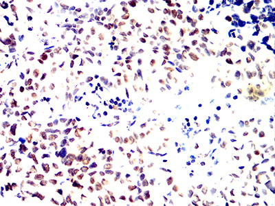
Figure 3:Immunohistochemical analysis of paraffin-embedded human liver tissues using RFA2 mouse mAb with DAB staining.
Immunohistochemical analysis
Figure 4:Immunohistochemical analysis of paraffin-embedded bladder cancer tissues using RFA2 mouse mAb with DAB staining.
Immunohistochemical analysis
Figure 5:Immunohistochemical analysis of paraffin-embedded esophageal cancer tissues using RFA2 mouse mAb with DAB staining.
Immunohistochemical analysis
Figure 6:Immunohistochemical analysis of paraffin-embedded rectum cancer tissues using RFA2 mouse mAb with DAB staining.
For Research Use Only. Not for use in diagnostic procedures.