PRKCG Primary Antibody

Item Information
Catalog #
Size
Price
Description

Protein kinase C (PKC) is a family of serine- and threonine-specific protein kinases that can be activated by calcium and second messenger diacylglycerol. PKC family members phosphorylate a wide variety of protein targets and are known to be involved in diverse cellular signaling pathways. PKC also serve as major receptors for phorbol esters, a class of tumor promoters. Each member of the PKC family has a specific expression profile and is believed to play distinct roles in cells. The protein encoded by this gene is one of the PKC family members. This protein kinase is expressed solely in the brain and spinal cord and its localization is restricted to neurons. It has been demonstrated that several neuronal functions, including long term potentiation (LTP) and long term depression (LTD), specifically require this kinase. Knockout studies in mice also suggest that this kinase may be involved in neuropathic pain development. Defects in this protein have been associated with neurodegenerative disorder spinocerebellar ataxia-14 (SCA14).

Product Overview
Entrez GenelD
5582
Aliases
PKCC; PKCG; SCA14; PKC-gamma
Clone#
1G5B3
Host / Isotype
Mouse / IgG1
Species Reactivity
Human
Immunogen
Purified recombinant fragment of human PRKCG (AA: 87-338) expressed in E. Coli.
Formulation
Purified antibody from tissue culture in PBS with 0.05% sodium azide
Storage
4°C; -20°C for long term storage
Product Applications
WB (Western Blot)
1/500 - 1/2000
ELISA
1/10000
References
1. Chem Biol Interact. 2010 Apr 15;185(1):25-32.
2. Biochem Biophys Res Commun. 2008 Aug 1;372(3):447-53.
Product Image
Western Blot
Figure 1: Western blot analysis using PRKCG mAb against human PRKCG (AA: 87-338) recombinant protein. (Expected MW is 54.4 kDa)
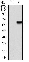
Western Blot
Figure 2: Western blot analysis using PRKCG mAb against HEK293 (1) and PRKCG (AA: 87-338)-hIgGFc transfected HEK293 (2) cell lysate.
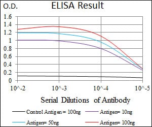
Elisa
Black line: Control Antigen (100 ng); Purple line: Antigen(10ng); Blue line: Antigen (50 ng); Red line: Antigen (100 ng);
For Research Use Only. Not for use in diagnostic procedures.