PPM1A Primary Antibody

Item Information
Catalog #
Size
Price
Description

The protein encoded by this gene is a member of the PP2C family of Ser/Thr protein phosphatases. PP2C family members are known to be negative regulators of cell stress response pathways. This phosphatase dephosphorylates, and negatively regulates the activities of, MAP kinases and MAP kinase kinases. It has been shown to inhibit the activation of p38 and JNK kinase cascades induced by environmental stresses. This phosphatase can also dephosphorylate cyclin-dependent kinases, and thus may be involved in cell cycle control. Overexpression of this phosphatase is reported to activate the expression of the tumor suppressor gene TP53/p53, which leads to G2/M cell cycle arrest and apoptosis. Three alternatively spliced transcript variants encoding distinct isoforms have been described.

Product Overview
Entrez GenelD
5494
Aliases
PP2CA; PP2Calpha; PP2C-ALPHA
Clone#
7F12H6
Host / Isotype
Mouse / IgG1
Species Reactivity
Human, Monkey
Immunogen
Purified recombinant fragment of human PPM1A (AA: 202-382) expressed in E. Coli.

Formulation
Purified antibody in PBS with 0.05% sodium azide
Storage
4°C; -20°C for long term storage
Product Applications
WB (Western Blot)
1/500 - 1/2000
FCM (Flow Cytometry)
1/200 - 1/400
ELISA
1/10000
References
1.Biol Psychiatry. 2011 Feb 15;69(4):360-5.
2.Cell Signal. 2009 Jan;21(1):95-102.
Product Image
Western Blot
Figure 1: Western blot analysis using PPM1A mAb against human PPM1A recombinant protein. (Expected MW is 45.9 kDa)
Western Blot
Figure 2: Western blot analysis using PPM1A mAb against HEK293 (1) and PPM1A (AA: 202-382)-hIgGFc transfected HEK293 (2) cell lysate.
Western Blot
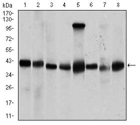
Figure 3: Western blot analysis using PPM1A mouse mAb against Jurkat (1), Jurkat (2), A431 (3), HeLa (4), HEK293 (5), Raji (6), MCF-7 (7), and COS7 (8) cell lysate.
Flow cytometric
Figure 4: Flow cytometric analysis of HeLa cells using PPM1A mouse mAb (green) and negative control (red).
Elisa
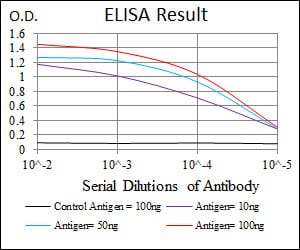
Black line: Control Antigen (100 ng); Purple line: Antigen(10ng); Blue line: Antigen (50 ng); Red line: Antigen (100 ng);
For Research Use Only. Not for use in diagnostic procedures.