MCM3

Item Information
Catalog #
Size
Price
Description

The protein encoded by this gene is one of the highly conserved mini-chromosome maintenance proteins (MCM) that are involved in the initiation of eukaryotic genome replication. The hexameric protein complex formed by MCM proteins is a key component of the pre-replication complex (pre_RC) and may be involved in the formation of replication forks and in the recruitment of other DNA replication related proteins. This protein is a subunit of the protein complex that consists of MCM2-7. It has been shown to interact directly with MCM5/CDC46. This protein also interacts with and is acetylated by MCM3AP, a chromatin-associated acetyltransferase. The acetylation of this protein inhibits the initiation of DNA replication and cell cycle progression. Several transcript variants encoding different isoforms have been found for this gene.

Product Overview
Entrez GenelD
4172
Aliases
HCC5; P1.h; RLFB; P1-MCM3
Clone#
1D4A3
Host / Isotype
Mouse / Mouse IgG1
Species Reactivity
Human, Monkey, Rat
Immunogen
Purified recombinant fragment of human MCM3 expressed in E. Coli.
Formulation
Purified antibody in PBS with 0.05% sodium azide
Storage
4℃; -20℃ for long term storage
Product Applications
WB (Western Blot)
1/500 - 1/2000
IHC_P(Immunohistochemistry)
1/200 - 1/1000
ICC (Immunocytochemistry)
1/50 - 1/200
FCM (Flow Cytometry)
1/200 - 1/400
References
1.Comput Math Methods Med. 2021 Oct 11;2021:8494260.
2.Biosci Rep. 2020 Jul 31;40(7):BSR20201503.
Product Image
Western Blot
Figure 1:Western blot analysis using MCM3 mouse mAb against PC-12 (1), HepG2 (2),Hela (3) and COS7 (4) cell lysate.
Immunohistochemical analysis
Figure 2:Immunofluorescence analysis of Hela cells using MCM3 mouse mAb (green). Blue: DRAQ5 fluorescent DNA dye. Red: Actin filaments have been labeled with Alexa Fluor- 555 phalloidin. Secondary antibody from Fisher (Cat#: 35503)
Immunofluorescence analysis
Figure 3:Flow cytometric analysis of Hela cells using MCM3 mouse mAb (green) and negative control (red).
Immunohistochemical analysis
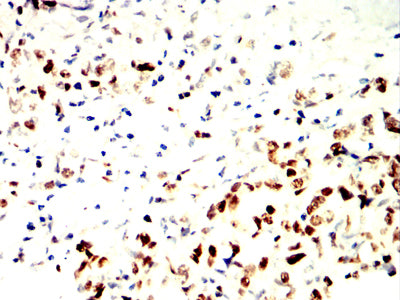
Figure 4:Immunohistochemical analysis of paraffin-embedded stomach cancer tissues using MCM3 mouse mAb with DAB staining.
Immunohistochemical analysis
Figure 5:Immunohistochemical analysis of paraffin-embedded rectum cancer tissues using MCM3 mouse mAb with DAB staining.
For Research Use Only. Not for use in diagnostic procedures.