KIT Primary Antibody

Item Information
Catalog #
Size
Price
Description

The c-Kit proto-oncogene is a member of the receptor tyrosine kinase family and, more specifically, is closely related to the platelet derived growth factor receptor (PDGFR). c-Kit, the normal cellular homolog of the HZ4-feline sarcoma virus transforming gene (v-Kit), encodes a transmembrane receptor. c-Kit regulates a variety of biological responses including chemotaxis, cell prolif- eration, apoptosis and adhesion. c-Kit is also identical with the product of the W locus in mice and, as such, is integral to the development of mast cells and hematopoiesis. The ligand for the c-Kit receptor (KL) has been identified and is encoded at the murine steel (SI) locus. Kit is the human homolog of the proto- oncogene c-Kit. Mutations in Kit are integral for tumor growth and progression in various cancers.

Product Overview
Entrez GenelD
3815
Aliases
PBT; SCFR; C-Kit; CD117
Clone#
1C5
Host / Isotype
Mouse / IgG1
Species Reactivity
Human
Immunogen
Purified recombinant fragment of human KIT expressed in E. Coli.
Formulation
Ascitic fluid containing 0.03% sodium azide.
Storage
4°C; -20°C for long term storage
Product Applications
WB (Western Blot)
1/500 - 1/2000
IHC_P(Immunohistochemistry)
1/200 - 1/1000
ELISA
1/10000
References
1. Curr Hematol Malig Rep. 2009 Apr;4(2):77-82.
2. Georgian Med News. 2010 Mar;(180):13-9.
Product Image
Western Blot
Figure 1: Western blot analysis using KIT mAb against human KIT (AA: 805-976) recombinant protein. (Expected MW is 45 kDa)
Western Blot
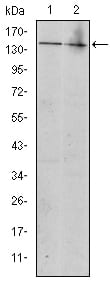
Figure 2: Western blot analysis using KIT mouse mAb against Jurkat (1) and Hela (2) cell lysate.
Immunohistochemical analysis
Figure 3: Immunohistochemical analysis of paraffin-embedded gastrointestinal stromal tumor using KIT mouse mAb with DAB staining.
Elisa
Red: Control Antigen (100ng); Purple: Antigen (10ng); Green: Antigen (50ng); Blue: Antigen (100ng);
For Research Use Only. Not for use in diagnostic procedures.