KHDRBS2 Primary Antibody
Item Information
Catalog #
Size
Price
Description
RNA-binding protein that plays a role in the regulation of alternative splicing and influences mRNA splice site selection and exon inclusion. Its phosphorylation by FYN inhibits its ability to regulate splice site selection. Induces an increased concentration-dependent incorporation of exon in CD44 pre-mRNA by direct binding to purine-rich exonic enhancer. May function as an adapter protein for Src kinases during mitosis. Binds both poly(A) and poly(U) homopolymers. Phosphorylation by PTK6 inhibits its RNA-binding ability (By similarity)
Product Overview
Entrez GenelD
202559
Aliases
SLM1; SLM-1; bA535F17.1
Clone#
3D12C11
Host / Isotype
Mouse / IgG1
Species Reactivity
Human, Mouse
Immunogen
Purified recombinant fragment of human KHDRBS2 (AA: 160-349) expressed in E. Coli.
Formulation
Purified antibody from tissue culture in PBS with 0.05% sodium azide
Storage
4°C; -20°C for long term storage
Product Applications
WB (Western Blot)
1/500 - 1/2000
FCM (Flow Cytometry)
1/200 - 1/400
ELISA
1/10000
References
1. Mol Biol Cell. 2003 Jan;14(1):274-87.
2.
2.
Product Image
Western Blot

Figure 1: Western blot analysis using KHDRBS2 mAb against human KHDRBS2 (AA: 160-349) recombinant protein. (Expected MW is 46.3 kDa)
Western Blot

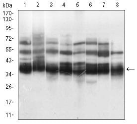
Figure 2: Western blot analysis using KHDRBS2 mouse mAb against K562 (1), HEK293 (2), NTERA-2 (3), Hela (4), HepG2 (5), Jurkat (6), A431 (7), NIH/3T3 (8) cell lysate.
Flow cytometric

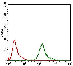
Figure 3: Flow cytometric analysis of K562 cells using KHDRBS2 mouse mAb (green) and negative control (red).
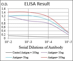
Elisa

Black line: Control Antigen (100 ng); Purple line: Antigen(10ng); Blue line: Antigen (50 ng); Red line: Antigen (100 ng);
For Research Use Only. Not for use in diagnostic procedures.

