Mouse Monoclonal Antibody to ACTH

Item Information
Catalog #
Size
Price
Description

This gene encodes a preproprotein that undergoes extensive, tissue-specific, post-translational processing via cleavage by subtilisin-like enzymes known as prohormone convertases. There are eight potential cleavage sites within the preproprotein and, depending on tissue type and the available convertases, processing may yield as many as ten biologically active peptides involved in diverse cellular functions. The encoded protein is synthesized mainly in corticotroph cells of the anterior pituitary where four cleavage sites are used; adrenocorticotrophin, essential for normal steroidogenesis and the maintenance of normal adrenal weight, and lipotropin beta are the major end products. In other tissues, including the hypothalamus, placenta, and epithelium, all cleavage sites may be used, giving rise to peptides with roles in pain and energy homeostasis, melanocyte stimulation, and immune modulation. These include several distinct melanotropins, lipotropins, and endorphins that are contained within the adrenocorticotrophin and beta-lipotropin peptides. The antimicrobial melanotropin alpha peptide exhibits antibacterial and antifungal activity. Mutations in this gene have been associated with early onset obesity, adrenal insufficiency, and red hair pigmentation. Alternatively spliced transcript variants encoding the same protein have been described.
Product Overview
Entrez GenelD
5443
Aliases
POMC; LPH; MSH; NPP; POC; CLIP; OBAIRH
Clone#
2B4A1
Host / Isotype
Mouse / IgG1
Immunogen
Purified recombinant fragment of human ACTH expressed in E. Coli.
Formulation
Purified antibody in PBS with 0.05% sodium azide
Storage
4℃; -20℃ for long term storage
Product Applications
WB (Western Blot)
1/500 - 1/2000
IHC_P(Immunohistochemistry)
1/200 - 1/1000
FCM (Flow Cytometry)
1/200 - 1/400
ELISA
1/10000
References
1.Front Neuroendocrinol. 2019 Jul;54:100773. 2.J Pediatr Endocrinol Metab. 2018 Jul 26;31(7):815-819.
Product Image
Elisa
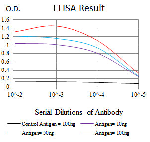
Figure 1:Black line: Control Antigen (100 ng);Purple line: Antigen (10ng); Blue line: Antigen (50 ng); Red line:Antigen (100 ng)
Western Blot
Figure 2:Western blot analysis using ACTH mAb against human ACTH recombinant protein. (Expected MW is 21.9 kDa)
Flow cytometric analysis
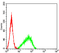
Figure 3:Flow cytometric analysis of K562 cells using ACTH mouse mAb (green) and negative control (red).
Immunohistochemical analysis
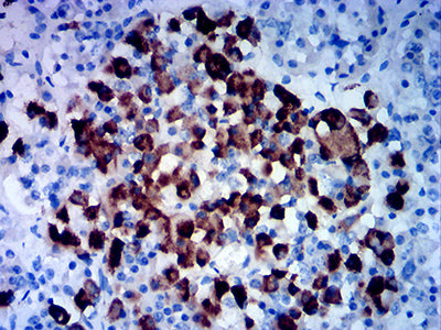
Figure 4:Immunohistochemical analysis of paraffin-embedded hypophysis tissues using ACTH mouse mAb with DAB staining.
For Research Use Only. Not for use in diagnostic procedures.